USMLE USMLE-STEP-1 Online Practice
Questions and Exam Preparation
USMLE-STEP-1 Exam Details
Exam Code
:USMLE-STEP-1
Exam Name
:United States Medical Licensing Step 1
Certification
:USMLE Certifications
Vendor
:USMLE
Total Questions
:847 Q&As
Last Updated
:May 25, 2026
USMLE USMLE-STEP-1 Online Questions &
Answers
Question 761:
A 22-year-old man recovers from a bout of hepatitis Aafter 3 weeks. One year after this infection, which of the following would a liver biopsy most likely demonstrate?
A. bridging fibrosis B. lymphocytes spilling out of the portal tracts with piecemeal necrosis C. lymphocytic infiltrates limited to portal triads D. massive hepatic necrosis E. normal histology
E. normal histology
Explanation
Section: Pathology and Path physiology Hepatitis A does not progress to a chronic phase so one year after infection the liver histology will appear as normal. The other choices are found in active hepatitis. Bridging fibrosis (choice A) and lymphocytes spilling out of the portal tracts with piecemeal necrosis (choice B) are major features of chronic hepatitis although both can be found in severe acute hepatitis. Lymphocytic infiltrates limited to portal triads (choice C) may be seen in mild acute or chronic hepatitis. Massive hepatic necrosis (choice D) is associated with fulminant hepatitis but can also be caused by various chemical and drug toxicities.
Question 762:
In cleaning the teeth in a patient, a dental hygienist accidentally cuts the gums of the posterior two molar teeth in the lower jaw on the lateral side. The pain of this injury is registered by which of the following nerves?
A. anterior, middle, and posterior superior alveolar nerves B. buccal nerve C. greater palatine nerve D. lingual nerve E. nasopalatine nerve
B. buccal nerve
Explanation
Section: Anatomy The gums on the lateral side of the mandibular molar teeth are innervated by the buccal nerve (long buccal nerve). All three superior alveolar nerves (choice A) supply the gums lateral to all maxillary teeth. The greater palatine nerve (choice C) innervates the gums medial to the maxillary premolar and molar teeth. The lingual nerve (choice D) supplies the gums medial to all mandibular teeth. The nasopalatine nerve (choice E) innervates the gums posterior to the maxillary incisors.
Question 763:
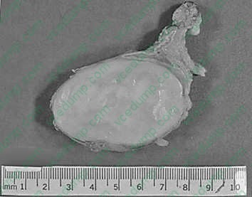
A 45-year-old woman complains of gradually increasing fatigue. On physical examination she is noted to have obesity, hypertension, buffalo hump deformity of her back, moon facies, abdominal striae, and muscle weakness. Radiographic imaging studies identify an abnormality of her right adrenal gland which is then surgically resected. The intact specimen weighed 24 g. A hemisection of the gland is displayed in below figure. What is the most likely diagnosis?
A. adrenal cortical adenoma B. Krukenberg tumor C. multifocal infarction D. neuroblastoma E. pheochromocytoma
A. adrenal cortical adenoma
Explanation
Section: Pathology and Path physiology The adrenal gland shown in figure contains a cortical adenoma. Grossly, these neoplasms appear as well- demarcated, round to oval, yellowish, solitary nodules arising within the adrenal cortex. The surrounding nonneoplastic cortex is thinned and atrophic. The underlying medulla is normal. The associated clinical findings suggest Cushing syndrome due to excessive secretion of cortisol by the adenoma. Krukenberg tumor (choice B) is an enlarged ovary due to metastatic carcinoma. Adrenal infarcts (choice C) may be associated with certain bacterial infections and shock. The gland appears diffusely hemorrhagic and necrotic, without the formation of a discrete tumor nodule. Neuroblastoma (choice D) is an adrenal tumor of infancy. The typical gross appearance is a large, tan hemorrhagic mass. Hypercortisolemia is not seen. Apheochromocytoma (choice E) appears as a hemorrhagic, red- tan medullary tumor. Clinically, there may be signs of excessive norepinephrine secretion.
Question 764:
On otoscopic examination, a patient is found to have a 2-cm mass protruding from a retraction pocket in his right tympanic membrane. The lesion is removed and determined to be a cystic mass lined by squamous epithelium containing desquamated cellular debris and a mononuclear infiltrate. Which of the following is the most likely cause of this lesion?
A. barotraumas B. chronic otitis media C. exostosis formation in the external auditory canal D. squamous cell carcinoma of the external auditory canal E. tympanosclerosis
B. chronic otitis media
Explanation
Section: Pathology and Path physiology A cholesteatoma is a cystic lesion lined by squamous epithelium and containing keratinaceous material. It is most probably formed by the protrusion of squamous epithelium from the middle ear canal through a perforation in the eardrum that was itself the result of chronic otitis media. Barotrauma (choice A) to the ear can be produced by sudden changes in atmospheric pressure compared to the relatively low middle ear pressure. This results in inflammation of the mucous membrane of the middle ear (serous otitis media). Exostosis formation in the external auditory canal (choice C) refers to the growth of a bony swelling that may occur with chronic exposure to cold water. Squamous cell carcinomas of the external auditory canal (choice D) are unusual and have a histological appearance similar to other squamous cell carcinomas (polygonal cells with prickles and pearls), which is quite different from the description given. Tympanosclerosis (choice E) of the tympanic membrane results from resolved acute otitis media producing acellular hyaline and calcific deposits in the tympanic membrane.
Question 765:
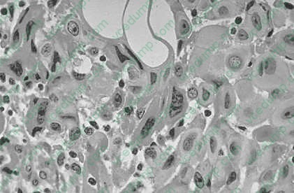
Aretroperitoneal mass was discovered during exploratory surgery on a 49-year-old woman. A photomicrograph of a section taken from the mass is shown in below figure. Which of the following terms would be most appropriate to describe its appearance?
A. anaplasia B. aplasia C. dysplasia D. hyperplasia E. metaplasia
A. anaplasia
Explanation
Section: Pathology and Path physiology The cells in figure show dramatic differences in the size, shape, and staining intensity of nuclei as well as differences in the cells overall, which allows one to say that they are pleomorphic. An additional factor is the lack of differentiation of these cells such that the cell type cannot be recognized, which is the definition of anaplasia. Aplasia (choice B) is a lack of growth of the anlage or primordium of an organ. Dysplasia (choice C) at the cellular level refers to disorderly maturation, usually of an epithelium.
Hyperplasia (choice D) is an increase in the number of cells and this process may be either physiological or pathological. Metaplasia (choice E) is the replacement of one mature cell type by another (e.g., columnar epithelium being replaced by squamous epithelium in a smoker's lung).
Question 766:
A 23-year-old female has serum electrolytes tested as part of a routine physical. The laboratory results reveal a mild degree of hypokalemia. Which of the following will promote movement of extracellular potassium into the intracellular fluid compartment and cause hypokalemia?
A. extracellular fluid hyperosmolality B. intravenous administration of a betaadrenergic blocker C. intravenous administration of insulin D. metabolic acidosis E. physical exercise
C. intravenous administration of insulin
Question 767:
Regarding the axon of the second-order neuron in the pathway for conscious awareness of fine, discriminative touch and vibratory sensation from the upper limb, which of the following is correct?
A. ascends the brainstem in the medial lemniscus B. decussates in the ventral white commissure of the spinal cord C. has its cell body in the nucleus gracilis D. is found in the dorsal funiculus of the spinal cord E. terminates in the nucleus cuneatus
A. ascends the brainstem in the medial lemniscus
Explanation
Section: Anatomy The sensations of discriminative touch and vibration are transmitted through the medial lemniscus. Pain and temperature pathways decussate in the ventral white commisssure (choice B). The nucleus gracilis (choice C) contains neurons that process sensory signals from the lower extremity. The second-order fibers carrying discriminative touch and vibration from the upper limb originate from neurons in the nucleus cuneatus (choice E). First order fibers are found in the dorsal funiculus (choice D).
Question 768:
Autoantibody testing of a patient with an autoimmune disorder reveals high titers of antibodies against the core proteins of small nuclear ribonucleoprotein particles (Smith antigen). Based on this finding, detection of which of the following is the most important initial clinical feature of this autoimmune disorder?
A. a lesion known as erythema chronicum migrans B. a rash in a butterfly distribution on the face C. antibody to single-stranded RNA D. carditis E. pneumonitis
B. a rash in a butterfly distribution on the face
Explanation
Section: Microbiology/Immunology The autoimmune disease described here by the presence of anti-Sm antibodies is Systemic lupus erythematosus (SLE). This autoimmune disease is initially recognized clinically by a rash on the face with a "wolf"-like shape and thus the name lupus. Such a butterfly rash (over the malar eminences of the face, hence a malar rash) is considered a distinctive manifestation of SLE. The development of erythema chronicum migrans is a pathognomonic feature of Lyme disease (choice A). Detection of antibodies to double-stranded DNA are pathognomonic for SLE (choice C). Arthritis, but not carditis (choice D) or pneumonitis (choice E), occurs frequently.
Question 769:
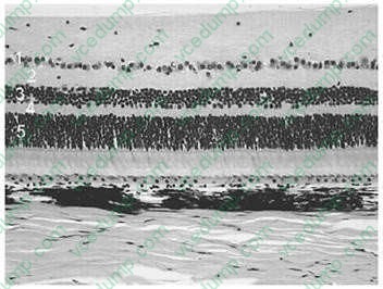
Retinitis pigmentosa is a hereditary disorder, which affects the photoreceptors (the rods and the cones) in the retina. These photoreceptors are located in which of the numbered layers in Figure below
A. 1 B. 2 C. 3 D. 4 E. 5
E. 5
Explanation
Section: Anatomy The retina contains three layers of cells. They are, from top to bottom, the ganglion cells (choice A), the bipolar cells (choice C), and the photoreceptor cells (rods and cones, choice E). The internal plexiform layer (choice B) contains the synapses between the bipolar cells and the ganglion cells. The external plexiform layer (choice D) contains the synapses between the photoreceptors and the bipolar cells. Remember that light enters from the top and traverses all the layers to reach the photoreceptors in the bottom layer.
Question 770:
An elderly patient with multi-infarct dementia also suffers from urinary incontinence and ataxia. Which of the following therapeutic procedures may be considered for this type of patient?
A. benzodiazepines B. electroconvulsive therapy C. megavitamin therapy D. urocholine E. ventriculoperitoneal shunt
E. ventriculoperitoneal shunt
Explanation
Section: Behavioral Science and Biostatics Normal pressure hydrocephalus is associated with the symptoms of dementia, ataxia, and urinary incontinence. On brain imaging, the ventricles are often enlarged, but cerebrospinal fluid (CSF) pressure is normal. Aventriculoperitoneal shunt is often therapeutic for this condition. Choices A, B, C, and D are inappropriate.
Nowadays, the certification exams become more and more important and required by more and more
enterprises when applying for a job. But how to prepare for the exam effectively? How to prepare
for the exam in a short time with less efforts? How to get a ideal result and how to find the
most reliable resources? Here on Vcedump.com, you will find all the answers.
Vcedump.com provide not only USMLE exam questions,
answers and explanations but also complete assistance on your exam preparation and certification
application. If you are confused on your USMLE-STEP-1 exam preparations
and USMLE certification application, do not hesitate to visit our
Vcedump.com to find your solutions here.