USMLE-STEP-1 Exam Details
-
Exam Code
:USMLE-STEP-1 -
Exam Name
:United States Medical Licensing Step 1 -
Certification
:USMLE Certifications -
Vendor
:USMLE -
Total Questions
:847 Q&As -
Last Updated
:May 25, 2026
USMLE USMLE-STEP-1 Online Questions & Answers
-
Question 291:
Which of the following quantitative comparisons of haloperidol (H) and olanzapine (O) is most accurate?
A. Option A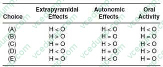
B. Option B
C. Option C
D. Option D
E. Option E -
Question 292:
Figure shows the quantal population doseresponse curves for the therapeutic and toxic effects of drugs X and Y. Both drugs are agonists at the same receptor to produce the therapeutic response, and the maximum responses obtained with each agent are the same. The toxicity curve in the figure shows the superimposed toxic response curves for drugs X and Y; they are identical in terms of the concentration dependence. Which of the following statements is most correct?
A. At 1 × 10⁻⁵ M both drugs cause adverse effects in 90100% of patients.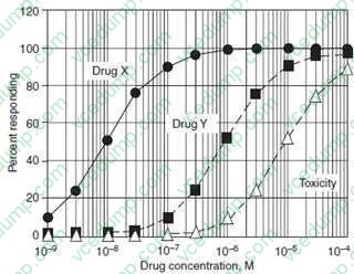
B. Drug X has a larger therapeutic index than drug Y.
C. Drug X is more efficacious than drug Y.
D. Drug Y is more potent than drug X.
E. Drug Y is safer than drug X. -
Question 293:
Clinical evidence indicates aspirin is effective in the control of numerous chronic conditions such as atherosclerosis. The principal cardiovascular benefit from aspirin is due to its ability to reduce the incidence and severity of thrombotic episodes. The anticoagulant effect of aspirin occurs through its ability to inhibit which of the following activities?
A. cyclooxygenase
B. fibrin cross-linking by factor XIIIa
C. phospholipase
D. thrombin binding to activated platelets
E. von Willebrand factor -
Question 294:
A young resident doing a fellowship in neuropathology is asked to review histological slides from the cerebral cortex of a 79-year-old nursing home resident, who died of multiinfarct dementia. The resident is asked to estimate the density of neurons in the infracted brain area. To prepare himself for the task, he first reviews slides from the normal areas of the cerebral cortex. Referring to following figure,which of the following structures does he correctly identify as neurons?
A. 1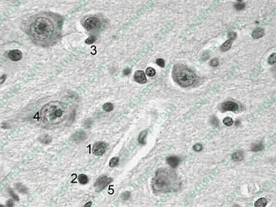
B. 2
C. 3
D. 4
E. 5 -
Question 295:
The structure indicated by arrow 1 in Fig. 1-2 is which of the following vessels?
A. brachiocephalic artery
B. left brachiocephalic vein
C. left common carotid artery
D. right brachiocephalic vein
E. superior vena cava -
Question 296:
Exhibit:
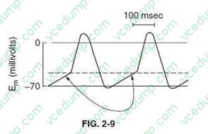
The action potentials shown in below figure represent those of which kind of cells?
A. cardiac nodal cells
B. myelinated motor axons
C. sensory neurons
D. skeletal muscle cells
E. ventricular Purkinje cells -
Question 297:
Numerous inherited disorders are the result of the expansion of trinucleotide (triplet) repeats either within the coding regions of genes or the untranslated regions of the resultant RNAs. Which of the following diseases has been shown to be caused by triplet expansion?
A. cystic fibrosis (CF)
B. Duchenne muscular dystrophy (DMD)
C. FH
D. Huntington disease (HD)
E. Menkes disease -
Question 298:
Which of the following correctly defines the term: p ?
A. equilibrium constant for the dissociation of HA to and
B. ion constant of water
C. negative log of the concentration of
D. pH at which a molecule is neutrally charged
E. pH at which an equivalent distribution of acid and conjugate base exist in solution -
Question 299:
Persistent fever and neutropenia with persistently negative blood cultures is often caused by which of the following?
A. fungi
B. gram-negative organisms
C. gram-positive organisms
D. viral infections -
Question 300:
If the RNA synthesized were translated in a eukaryotic in vitro translation system, what would be the composition of the resultant peptide?
A. C-T-I-P-Y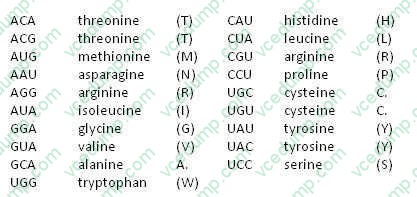
B. H-S-I-A-C
C. M-L-W-N
D. T-C-Y-G-M
E. V-R-Y-R-T
Tips on How to Prepare for the Exams
Nowadays, the certification exams become more and more important and required by more and more enterprises when applying for a job. But how to prepare for the exam effectively? How to prepare for the exam in a short time with less efforts? How to get a ideal result and how to find the most reliable resources? Here on Vcedump.com, you will find all the answers. Vcedump.com provide not only USMLE exam questions, answers and explanations but also complete assistance on your exam preparation and certification application. If you are confused on your USMLE-STEP-1 exam preparations and USMLE certification application, do not hesitate to visit our Vcedump.com to find your solutions here.