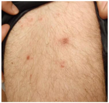
A 24-year-old man comes to the office because of a 2-day history of a red, itchy rash on his buttocks and legs. Four days ago, he returned from a cruise to the Caribbean, during which he swam in the ship's pool and used the hot tub. He appears well. His vital signs are within normal limits. Physical examination shows the findings in the photograph. The infectious agent causing these findings most likely began to proliferate in which of the following locations?
A. Apocrine gland
B. Dermis
C. Eccrine gland
D. Hair follicle
E. Sebaceous gland
Correct Answer: D
Question 582:
A 14-year-old girl is brought to the physician after her mother learned that she began having sexual intercourse with various partners 1 month ago. She does not use condoms or other contraception. The mother is concerned about her behavior. The patient's parents separated 3 months ago. She had been an honor student and excelled in sports and leadership positions at school before the separation. Since the separation, however, she has become sullen, defiant, and rebellious. She has begun smoking cigarettes, disobeying her curfew, and being truant from school. This patient is most likely using which of the following defense mechanisms?
A. Acting out
B. Displacement
C. Projection
D. Reaction formation
E. Sublimation
Correct Answer: A
Question 583:
A 45-year-old man with cirrhosis due to alpha1-antitrypsin deficiency receives a liver transplant. Although currently in good health, he is at increased risk of developing which of the following types of emphysema?
A. Centriacinar
B. Compensatory
C. Interstitial
D. Panacinar
E. Paraseptal
Correct Answer: D
Explanation:
There are two main morphologic forms of emphysema, centriacinar and panacinar. The panacinar variant is related to alpha1-antitrypsin deficiency; the entire acinus is enlarged, from the respiratory bronchiole to the distal alveoli. Centriacinar emphysema is characterized by enlargement of the central portions of the acinus, i.e., the respiratory bronchiole, and its pathogenesis is related to exposure to tobacco products and coal dust. Interstitial emphysema is not a true form of emphysema. It results from penetration of air into the pulmonary interstitium. This may occur when alveolar tears develop because of a combination of coughing and airway obstruction (e.g., children with whooping cough) or a chest wound that injures the underlying lung parenchyma (e.g., a fractured rib). Compensatory emphysema and paraseptal emphysema are associated with scarring. Both are frequent but usually clinically silent. Paraseptal emphysema, however, may lead to spontaneous pneumothorax in young patients. In fact, this form is more severe in areas adjacent to the pleura, where large, cyst-like structures may develop and rupture into the pleural cavity.
Question 584:
A 12-year-old girl has a temperature of 102.5 F and a sore throat. Two days later, she develops a diffuse erythematous rash and is taken to her pediatrician. On physical examination, there is circumoral pallor, and an erythematous rash with areas of desquamation is noted. The myocardial damage that can follow this infection is produced in a manner similar to the damage associated with which of the following disorders?
A. Atopic allergy
B. Systemic lupus erythematosus
C. Contact dermatitis
D. Graft-vs-host disease
E. Graves disease
F. Idiopathic thrombocytopenic purpura
G. Myasthenia gravis
H. Rheumatoid arthritis
I. Serum sickness
Correct Answer: F
Explanation:
This is a case of rheumatic fever, which is an immunologically mediated sequela to Streptococcus pyogenes pharyngitis. It is a type II cytotoxic hypersensitivity, involving antibodies that bind to cardiac tissue, activate complement, and thereby cause cell destruction. It is therefore most similar to idiopathic thrombocytopenic purpura, which is also a form of type II cytotoxic hypersensitivity, in this case mediated by antibodies against platelets producing complement fixation and causing the clotting dyscrasia. Atopic allergy is a form of type I hypersensitivity, mediated by IgE antibodies and basophils and mast cells. Contact dermatitis is a form of type IV hypersensitivity mediated by T cells and macrophages. Graft-vshost disease is a form of type IV hypersensitivity mediated by T cells and macrophages. Graves disease is a form of type II hypersensitivity, but it is NOT cytotoxic in its action. Instead, antibodies to the TSH receptors on thyroid cells cause overstimulation of the gland and its eventual exhaustion. Myasthenia gravis is a form of type II hypersensitivity, but NOT of the cytotoxic variety. In this case, antibodies to the acetylcholine receptors on neurons diminish neurotransmission. Rheumatoid arthritis is a form of type III hypersensitivity, caused by immune complex deposition in joints and subsequent activation of complement. Serum sickness is a form of type III hypersensitivity, caused by immune complex deposition. Systemic lupus erythematosus is a form of type III hypersensitivity, caused by immune complex deposition.
Question 585:
A 24-year-old woman in her third trimester of pregnancy presents with urinary frequency and burning for the past few days. She denies fever, nausea, vomiting, or chills. She takes no medications besides prenatal vitamins and is generally in good health. Physical examination is remarkable for mild suprapubic tenderness, and a urine dipstick is positive for white blood cells, protein, and a small amount of blood. Culture produces greater than 100,000 colonies of gram-negative bacilli. Which of the following attributes of this uropathogenic organism is most strongly associated with its virulence?
A. Bundle-forming pili
B. GVVPQ fimbriae
C. Heat labile toxins
D. Heat stable toxins
E. P pili
F. Type 1 pili
Correct Answer: E
Explanation:
Urinary tract infections are the most common bacterial infections encountered during pregnancy, and Escherichia coli is the most commonly isolated organism. In the U.S., 70% of cases are caused by P pilipositive strains. Bundle-forming pili are found in enteroaggregative E. coli (EAEC).GVVPQ fimbriae are found in EAEC. Heat labile toxins are pathogenic factors in enterotoxic strains (ETEC). Heat stable toxins are pathogenic factors in ETEC or EAEC. Type 1 pili are a major pathogenic factor in ETEC.
Question 586:
A 15-year-old is brought to the emergency department in a coma. An alert ambulance attendant notes that the patient's breath smells like acetone. This observation is most consistent with which of the following diagnoses?
A. Alcohol intoxication
B. Diabetic hyperosmolar coma
C. Diabetic ketoacidosis
D. Heroin overdose
E. Profound hypoglycemia
Correct Answer: C
Explanation:
The smell of acetone on the breath of a comatose patient is an important, rapid diagnostic clue that strongly suggests ketoacidosis and is usually seen in patients with poorly controlled type 1 diabetes. Other features of diabetic ketoacidosis include high blood glucose, increased serum osmolality, hypovolemia, acidosis, and electrolyte imbalance. In alcohol intoxication, the breath will smell like alcohol. Diabetic hyperosmolar coma usually is seen in older patients with type 2 diabetes and is not characterized by ketoacidosis. Since there is no acetone production, there is no specific scent to the breath. In heroin overdose, no acetone production occurs and there is no specific scent to the breath. In hypoglycemic coma, which can occur in diabetics with insulin overdose, no acetone production occurs and there is no specific scent to the breath.
Question 587:
A 65 year-old man is admitted to the coronary care unit with a diagnosis of a large myocardial infarct (MI) of the left ventricle. On his 6th postinfarct day, he goes into shock and dies, manifesting signs and symptoms of cardiac tamponade. Which of the following complications is the most likely cause of this patient's death?
A. Aortic dissection
B. Extension of previous MI
C. Fatal arrhythmia
D. Rupture of the left ventricular wall
E. Rupture of papillary muscle
Correct Answer: D
Explanation:
Rupture of the free left ventricular wall is a frequently fatal complication that may occur in the first week after myocardial infarction (MI). At this stage, the infarcted area is composed of friable necrotic myocardium and early granulation tissue. It is during this crucial phase, therefore, that rupture usually occurs. Blood rushes out, filling the pericardial sac and causing compression of the left ventricle. Cardiac tamponade ensues, and the patient usually dies of acute cardiogenic shock. Aortic dissection is not a complication of MI, although cardiac tamponade may also follow this acute condition when dissection works its way back toward the aortic root. Aortic dissection usually develops in aortas affected by cystic medial degeneration (CMD), which is due to fragmentation of elastic laminae with accumulation of myxoid material in the aortic media. CMD may be either sporadic or associated with Marfan syndrome. Extension of a previous MI may occur in the first few hours or days after MI. It may aggravate or precipitate cardiogenic shock and/or arrhythmias, but it does not cause cardiac tamponade. Arrhythmias are frequent complications of MI and are often fatal, producing cardiac arrest (ventricular fibrillation) or aggravating cardiac dysfunction. If infarction involves papillary muscles, these may rupture. This complication is followed by valvular dysfunction and may manifest with signs of mitral regurgitation and acute congestive heart failure.
Question 588:
A 14-year-old boy has just moved with his family from Brazil to the U.S. He starts complaining of shortness of breath and palpitations. Chest x-ray films demonstrate pulmonary congestion, and EKG shows alterations in heart rhythm. Echocardiography reveals biventricular dilatation with massive cardiac enlargement. An endomyocardial biopsy shows diffuse interstitial fibrosis, myocyte necrosis, chronic inflammation, and the presence of intracellular protozoan parasites. The patient may also develop which of the following complications?
A. Achalasia
B. Splenomegaly
C. Chronic arthritis
D. Cysts in the brain
E. Pleuritis
Correct Answer: A
Explanation:
The patient has myocarditis due to Trypanosoma cruzi. This infectious condition, known as Chagas disease, is endemic in vast areas of South America and is transmitted from person to person by triatomids known as “kissing bugs.” Experts assess the number of persons with Chagas disease at about 7 million, with about 35 million at risk in South America. T. cruzi is an intracellular protozoon that localizes mainly in the heart and nerve cells of the myenteric plexus, leading to myocarditis and dysmotility of hollow organs, such the esophagus, colon, and ureter. Cardiac involvement manifests with ventricular dilatation and congestive heart failure secondary to myocyte necrosis and fibrosis. Intracellular parasites can be visualized in tissue sections. Chagas disease is a cause of acquired achalasia, in which the distal third of the esophagus dilates because of loss of its intrinsic innervation. A similar pathologic mechanism accounts for megacolon and megaureter in Chagas disease. The remaining choices refer to different infectious conditions that may also involve the myocardium: Chronic arthritis is a manifestation of the chronic stage of Lyme disease, which is caused by Borrelia burgdorferi and is transmitted to humans by deer ticks. Skin, CNS, and heart are the main targets of this infection. Cysts in the brain (cysticerci) may develop as a consequence of infestation by the tapeworm Taenia solium. Humans acquire this parasite by ingesting the eggs from undercooked pork. Cysticercosis may also affect the heart, skeletal muscle, and skin. Group B coxsackievirus infections cause pleuritis and myocarditis, manifesting with fever, chest pain, and, if myocarditis is severe, congestive heart failure. As in any form of viral myocarditis, the myocardium is infiltrated by lymphocytes, but there are no morphologic markers specific for Coxsackievirus infection. Splenomegaly, often of massive proportions, is seen in patients with malaria. Plasmodium organisms can also invade the myocardium, leading to myocarditis.
Question 589:
A patient arrives in the emergency department after having been stabbed. He has sustained a penetrating wound in the left fourth intercostal space immediately lateral to the sternal border. Which of the following thoracic structures is most likely to have been injured?
A. Left atrium
B. Left ventricle
C. Right atrium
D. Right ventricle
E. Upper lobe of the left lung
Correct Answer: D
Explanation:
The right ventricle forms most of the anterior wall of the heart and extends from approximately the right border of the sternum to approximately 2 inches to the left of the sternum at the level of the fourth intercostal space. The left atrium forms the posterior wall of the heart. The only portion of the left atrium seen on the anterior surface of the heart is the left auricular appendage, which is at the level of the second intercostal space on the left. The left ventricle forms most of the left border of the heart and the diaphragmatic surface of the heart. It forms the anterior wall of the heart in a region from approximately 2-3 inches from the left border of the sternum from the third to the fifth intercostal space. The right atrium forms the right border of the heart. Its anterior surface is on the right side of the sternum from approximately the third rib to the sixth rib. The left lung is displaced away from the sternum on the left side by the presence of the heart.
Question 590:
A 68-year-old man sustains a myocardial infarct resulting from thrombotic occlusion at the origin of the left circumflex artery. Cardiac catheterization demonstrates that the patient has a left dominant coronary circulation. In which of the following areas of the heart has ischemic necrosis most likely occurred?
A. Apex of left ventricle and anterior portion of septum
B. Lateral left ventricular wall and posterior portion of the septum
C. Lateral wall of the left ventricle only
D. Posterior portion of the septum only
E. Right ventricular wall
Correct Answer: B
Explanation:
A right dominant coronary circulation is present when the posterior descending branch originates from the right coronary artery (80% of individuals). On the contrary, the posterior descending artery originates from the left circumflex artery in a left dominant circulation (20% of individuals). The posterior descending branch gives blood to the posterior half of the interventricular septum. Occlusion of the left circumflex artery in a left dominant circulation will therefore lead to ischemic necrosis in the left ventricular wall and the posterior interventricular septum. The apex of the left ventricle is dependent on the anterior descending branch; thus, occlusion of the left circumflex does not affect this portion of the left ventricle. Infarction of the lateral (free) wall alone will result from occlusion of the circumflex in a right dominant circulation. An isolated infarct of the posterior interventricular septum arises from occlusion of the posterior descending branch. Isolated infarcts of the right ventricular wall are very rare and would be caused by occlusion of branches of the right coronary artery.
Nowadays, the certification exams become more and more important and required by more and more enterprises when applying for a job. But how to prepare for the exam effectively? How to prepare for the exam in a short time with less efforts? How to get a ideal result and how to find the most reliable resources? Here on Vcedump.com, you will find all the answers. Vcedump.com provide not only USMLE exam questions, answers and explanations but also complete assistance on your exam preparation and certification application. If you are confused on your USMLE exam preparations and USMLE certification application, do not hesitate to visit our Vcedump.com to find your solutions here.